Here is a description of one brain injury lawsuit successfully prosecuted by Patrick Malone of Stein,
Mitchell & Mezines. This lawsuit concerned medical malpractice by a radiologist and by a
neurologist in failing to prevent a large and debilitating stroke suffered by a client of our firm.
This story shows some of the key features involved in successfully developing and prosecuting
a brain injury lawsuit.
Sharon Burke v. Groover Christie & Merritt
Introduction
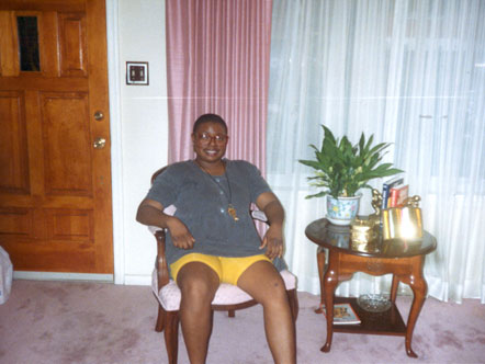
In December 1999, Sharon Burke was a successful retail men's wear store manager in the
Washington suburbs of Prince George's County, Maryland. She was 40 years old, single, and
popular with her friends for her quick wit and positive outlook.
Sharon Burke two years before her stroke:
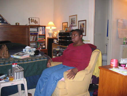
Sharon after the stroke:
In December 1999, Sharon Burke underwent an MRI scan of the brain because she had started
experiencing strange symptoms: numbness and tingling of his arms and legs, mostly on one side.
The scan was reported by radiologists at Groover Christie & Merritt, a large Washington-based
radiology group practice, as showing signs of multiple sclerosis, a degenerative disease where
nerves lose their insulation sheathing.
The symptoms went away, and Ms. Burke's neurologist did nothing until she came back to him
in July 2000, seven months later, complaining of similar symptoms. This time the symptoms
were more dramatic: she had had episodes where her legs suddenly gave way and she fell.
Another MRI scan was ordered. This time the report was more equivocal. The radiologist said
the scan looked like Ms. Burke might have multiple sclerosis, or an inflammation of blood
vessels in her brain, or a stroke from a blood clot.
An MRI scan of the brain was misread by a radiologist
That report was a misreading of what was on Sharon Burke's brain scan. The July scan actually
showed a blockage of one of the major blood vessels feeding the brain � the right internal carotid
artery � and clear evidence of stroke damage in the parts of the brain fed by that artery. Sharon
Burke went on to suffer a major stroke several months later, a stroke that could have been
prevented if her scan in July 2000 had been read correctly and she had been put on blood-thinning
medications to block the development of the clots that damaged her brain.
The incorrect brain scan report by Dr. William Higgins of Groover Christie misled both Dr. Stuart
Goodman, Ms. Burke's neurologist at the time of the scan, and another neurologist whom Ms.
Burke's family took her to in September 2000, frustrated by Dr. Goodman's failure to figure out
what was wrong with her. Both neurologists ordered a series of tests to chase down the possibility
of multiple sclerosis and other diseases which Ms. Burke didn't have. Ironically, the second
neurologist looked at Ms. Burke's brain scans himself just a few days before she suffered a major
stroke and had scheduled her to have imaging of the blood vessels in the brain � but the orders
came too late to get the tests done before her stroke.
On the morning of October 23, 2000, Ms. Burke's mother found her in her bedroom, unable to
speak or move. She was taken to a hospital, where imaging tests found the blood clots in her neck
arteries and the arteries in the brain. But it was too late to prevent major brain damage.
After Ms. Burke had gone through months of rehabilitation, and she had recovered only a fraction
of her pre-stroke abilities, her mother, Wilhelmina Torian, brought her to the lawyers at Stein,
Mitchell & Mezines seeking answers about whether the doctors had dropped the ball in their
care. The lawyers obtained all the relevant medical records and copies of the two MRI scans done
before the major stroke, as well as post-stroke brain imaging. These were organized and sent to
independent experts in the fields of neuroradiology (radiology that focuses on the brain and
spinal cord) and neurology. These experts reported back that both neurologists as well as the
radiology group were negligent in their care of Ms. Burke and had caused her to suffer an unnecessary
stroke.
Suit was filed late in 2001. Depositions were taken of both neurologists, the radiologists involved in
the two pre-stroke scans, and expert witnesses hired by both sides. Because of various delays in the
court system, the case did not reach trial until March 2004. Trial took place in the Superior Court of
the District of Columbia, Judge Mary Terrell presiding.
Here is some of the key evidence that Ms. Burke's lawyers used to prove their case. This first chart
shows a portion of the brain's blood supply (visual pop-up): the carotid arteries leading up from the front of the neck
and the cerebral arteries that flow from the carotids. In the side view on the right, you can see that
the carotid artery makes an S-curve as it comes into the base of the brain. It was at this curved
section that Ms. Burke's carotid artery was shown to be blocked in the July 2000 scan.
Here is a chart comparing the December 1999 MRI scan to the July 2000 scan. These are both
horizontal slices through the lower part of the brain. At the top of each scan, you can see the eyes
and the nasal cavities. The boxed area just behind the nasal cavities is the spinal cord with a
double-barreled cut through the carotid arteries on each side. On the December scan, the normal
flowing blood vessel appears as two black holes on each side of the spinal cord. On the July scan,
the right internal carotid artery now appears as two white holes instead of black.
This was never reported by the Groover Christie radiologist, who testified at trial that he did not do
so because he thought it was an "artifact" and not a true blockage, and he didn't want to confuse
the neurologist. Ironically, he did confuse two separate neurologists, who both testified that the
information about a blocked carotid artery would have completely changed their thinking about what
was wrong with Ms. Burke. A blocked major vessel does not go with either multiple sclerosis or
blood vessel inflammation, the other two alternate diagnoses being considered.
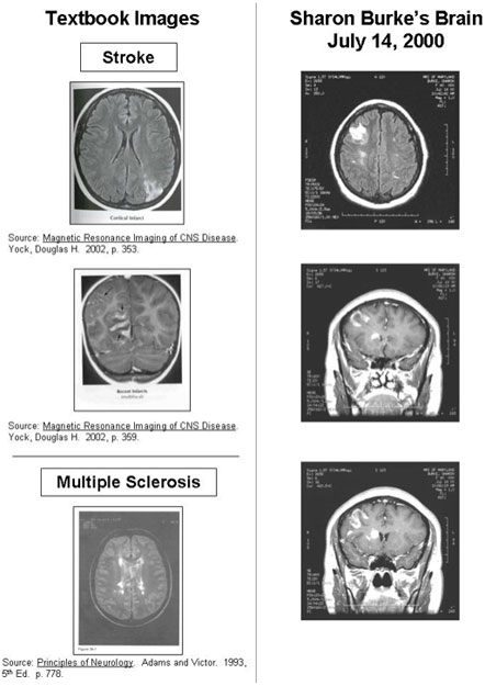
There were other reasons why the July 2000 MRI scan should have been reported as showing stroke
and not other possibilities. This was illustrated by comparing textbook images of stroke and multiple
sclerosis to what was seen in Ms. Burke's brain on other images from the same scan:
The areas of white seen in the upper left (on the patient's right side) in Ms. Burke's images showed a
wedge-shaped pattern (top image) and gyriform appearance (lower two images) � a curving snake-like
pattern that means the surface of the brain's cortex has been damaged. These are both indicative of
stroke and are completely unlike the deep damage near the brain's ventricles seen in multiple sclerosis.
Ms. Burke's case against the radiology group was supported by the testimony of David Yousem, M.D.,
chief of neuroradiology at Johns Hopkins Hospital in Baltimore. Dr. Yousem testified that Dr. Higgins'
official report violated the "standard of care" for neuroradiologists in five distinct ways. First, he failed
to report a major abnormality, the blocked carotid artery. Second, Dr. Higgins gave in his official report
a confusing list of three possible "differential diagnoses" for the abnormalities in Ms. Burke's scan: multiple
sclerosis, vasculitis (or arteritis) and ischemia, and he listed them in that order, leading both neurologists
who ultimately relied on this report to the natural conclusion that multiple sclerosis was the most likely
cause of the changes in her scan. Third, Dr. Higgins failed to compare this scan to her prior MRI scan
from December 1999, which would have revealed that the blocked carotid artery was a new finding and
thus no "artifact." Fourth, Dr. Higgins failed to recommend follow-up studies to determine the significance
of the blocked carotid artery. Fifth, Dr. Higgins failed to call the treating neurologist and alert him that
the diagnosis had changed, from the "multiple sclerosis" that Goodman had put on the request slip for
the MRI, to stroke caused by a blood clot in the right internal carotid artery.
Dr. Yousem's testimony was backed by references to published standards then in effect from the American
College of Radiology, the national professional society of radiologists. (Read the ACR 2000 standards for
reporting diagnostic radiology findings by clicking here. )
Ms. Burke's treating neurologist also failed to provide competent care.
Dr. Goodman, Ms. Burke's neurologist who ordered the July 2000 brain scan, was also criticized by expert
independent neurologists at the trial. Ms. Burke's symptoms reported to him were classic for "transient
ischemic attacks," which are small strokes that can act as warning signs of big strokes to come. He failed
to pursue a rational plan to determine what was wrong with her. The complex series of events that led to
Ms. Burke's preventable stroke in October 2000 were summarized in a timeline chart reproduced here (visual pop-up).
The two tests highlighted in yellow at the lower right of the timeline, both done just after her major stroke,
could have prevented the stroke if done earlier because they would have led to the correct diagnosis.
According to the neurologists who testified on Ms. Burke's behalf, Dr. Goodman had several clues that
should have led him to do these tests in spite of the confusing report received from the radiologist concerning
the July brain scan.
The testimony about Ms. Burke's injuries and damages
The trial featured extensive testimony about what the October 2000 stroke had done to Ms. Burke. The
earlier small strokes had left her functioning largely intact. This final stroke, however, devastated her
brain because it occurred on her dominant left side of the brain and involved areas important for thinking
and speech.
According to the testimony of Dr. Daniel Weinberger, a renowned behavioral neurologist who examined
Ms. Burke and reviewed reports of neuropsychological testing done on her, Ms. Burke, who was 44 years
old at the time of trial, has a vascular dementia that has rendered her brain less functional cognitively
than that of an 80-year-old person. She has "psychomotor slowing" that renders her slow in all activities.
Ms. Burke lost 20-25% of her IQ, has a reading comprehension at the third or fourth grade level plus
an "expressive aphasia" that renders her speech halting and difficult. She is easily confused and is
vulnerable to being taken advantage of. She has serious problems with short-term memory. Even on
the neuropsychological examination done for the defense lawyers, she failed approximately 55 of the 60
tests given.
Ms. Burke manages to survive on a day-to-day basis with five to six daily telephone calls to her mother,
plus multiple visits each week from her mother. Her mother carries all heavy objects back and forth from
one level of Ms. Burke's townhouse to the other, because Ms. Burke's residual right-sided weakness
prevents her from carrying objects on stairs. Mrs. Torian also does all the heavy cleaning. Ms. Burke
suffers daily from "cognitive exhaustion" due to the great efforts she must make mentally just to perform
tasks that normal people can do on "autopilot," according to BeverlyWhitlock, head of the Head Injury
Rehabilitation and Referral Services in Rockville, Md., (http://www.headinjuryrehab.org) where Ms.
Burke has had therapy for two years. Thus Ms. Burke needs to take a nap after a four-hour workday
at her volunteer "sheltered" job at Prince George's Hospital Center, where she does minor filing tasks
and document shredding. Ms. Burke needs close help with all "executive tasks" that require planning.
A trip to the grocery store is a day-long endeavor, according to her mother, because Ms. Burke forgets
what she needs, forgets where things are in the store, and exercises poor judgment about the quantities
she should purchase.
Ms. Burke's social life has been destroyed. She is acutely self-conscious about her inability to speak normally
and so avoids contacts with outsiders. Her entire social life is her mother, plus her sister's occasional visits
from North Carolina. She occupies herself with computer games and television where she formerly had a
busy social life with friends.
It was undisputed at trial that Ms. Burke's career in retail sales management was ruined. The defense
presented non-credible vocational testimony that she might be able to work as a telephone answering
person, a job that all other witnesses said would be preposterous for her because of her word-finding
difficulty related to the stroke, a condition called "expressive aphasia."
Ms. Burke will need to go into an assisted living facility because of her inability to manage her own affairs.
Her mother was 71 years old at the time of trial in March 2004, and the plaintiff's "life care plan" predicted
she would need to make the transition to assisted living in approximately six years. This plan was fully
supported by the objective evidence of Ms. Burke's cognitive and physical disabilities. The plan was
endorsed by Dr. Weinberger and by Beverly Whitlock, the head of the brain injury rehabilitation facility
which Ms. Burke has attended since 2002.
The defense called several witnesses who were hired to examine Ms. Burke and who tried to minimize the
damage that Ms. Burke had suffered. None of these persons had an M.D. Their credibility was impeached
by cross-examination showing that they had failed to interview significant persons such as Ms. Burke's
mother and her therapists, and because of their failure to fully grasp all of Ms. Burke's disabilities.
After two days of deliberations, the jury, which was dominated by young professionals, awarded $5.7
million in damages to Ms. Burke. Included were specific sums to pay her back for the lost lifetime wages
from her retail sales management career, other money to fund her future care needs, and money for her
lost enjoyment of life.
Legal Consultation
The lawyers at Stein, Mitchell & Mezines provide free and confidential consultations to help sort out your legal options. Contact us with this form to provide basic information to get the process started: